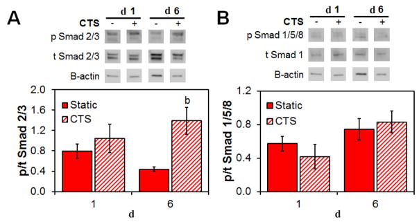
Fig. 6.
Differences in the activation of canonical vs. non-canonical Smad pathways for MSCs within the CG scaffold in response to CTS. Relative phosphorylation of (A) the non-canonical Smad 2/3 and (B) the canonical Smad 1/5/8 at day 1 and 6 as determined by immunoblot. Data are represented as mean ± SEM (n = 3). b Significantly higher expression (p < 0.05) than static group at same day.

