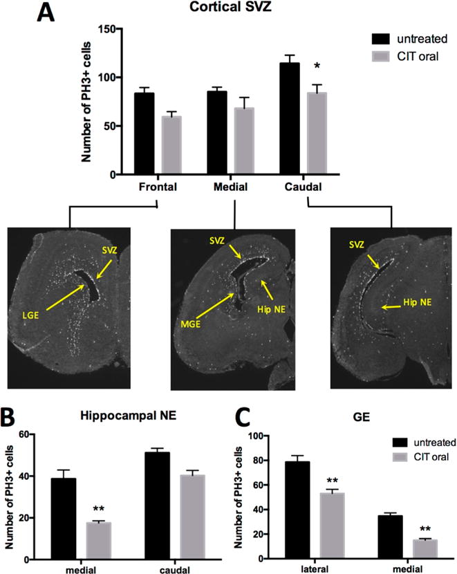
Figure 3.

Effect of in utero exposure to CIT on fetal brain neurogenesis at GD17. (A) Immunohistochemistry shows a significant decrease in the mean number of cells staining positive for PH3 in the caudal sections of the fetal cortical SVZ after chronic exposure of timed-pregnant mice to CIT in drinking water: SVZ = cortical subventricular zone (*p = 0.0382 versus untreated). (B) The medial part of the fetal hippocampal neuroepithelium (Hip NE) showed a significant decrease in mean number of PH3+ cells in maternal CIT-exposed embryos (**p = 0.0012 vs untreated). (C) Chronic maternal CIT exposure results in a significant decrease in the mean number of PH3+ cells in the lateral and medial fetal ganglionic eminence (GE) in comparison to untreated (**p = 0.0015 and p = 0.0086, respectively). All comparisons were made by two-way ANOVA with Bonferroni correction. Each column represents mean ± SEM. Results were obtained from 1 embryo per dam with n = 4 dams for control and n = 3 dams for treatment.
