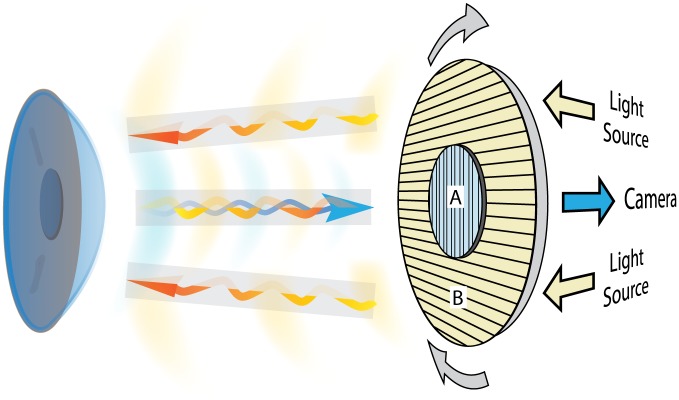
Fig 1. Schematic representation.
Principle of polarimetric interferometry of the corneal stroma. The incident light projected by the LED light source passes through the first polarized filter (B) and illuminates the cornea. The backscattered light after interaction with corneal tissue passes through the second cross-polarized filter (A) and is captured by the detector camera.

