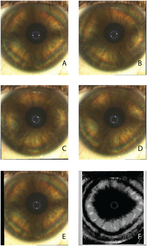
Fig 4. Corneal polarimetric interferometry of type A pattern.
Frames of representative raw sequence (A-E) in a case in which the isogyre maintains its corneal cross-shaped pattern with the rotating scan of the LCP. (F) The final summary static image (SUM image) is characterized by a single “pear shaped” dark area with the major axis along the horizontal meridians. Peripheral isochromes are visible on individual photograms.

