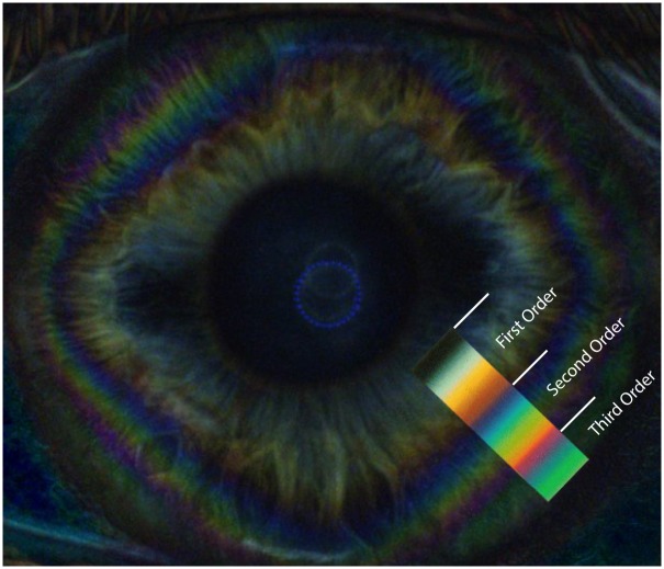
Fig 7. Corneal isochromatic interference figures in polarimetric interferometry enhanced after subtraction of background image.
Colored image showing the peripheral corneal isochromes, with a rounded square morphology. The matching changes of colour bands of the 1st through 3rd order of the Michel-Levy colour sequence are shown in the colour overlay.

