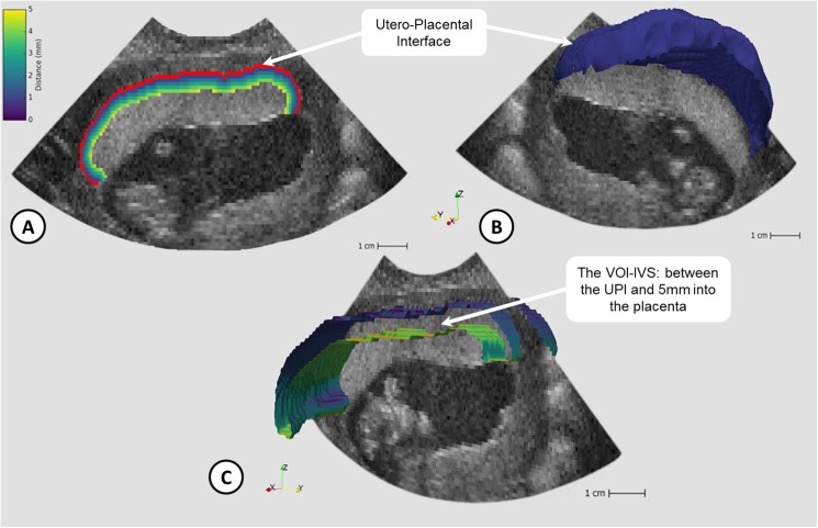
Fig 1. Visualisation of utero-placenta interface (UPI) and volume of interest (VOI-IVS: 3D ‘slice’ of tissue between the UPI and 5mm into the placenta containing the intervillous space (IVS)).
Fig 1a) 2D B-Mode image with UPI overlaid (red) and voxels up to 5mm into placenta labelled based on distance; Fig 1b) 3D UPI (blue) shown with 2D B-mode slice; Fig 1c) Section of the 3D VOI-IVS coloured by distance away from UPI.

