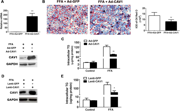
Fig 3. CAV1 overexpression ameliorated fat accumulation in L02 cells and AML12 cells induced by FFA.
CAV1 was overexpressed using the adenovirus recombination system via a transient transfection in L02 cells and the lentivirus-constructed stable transfection system in AML12 cells. (A-C) L02 cells were transfected with an adenovirus recombination plasmid containing full-length human CAV1 DNA to overexpress CAV1 (Ad-CAV1); an adenovirus empty vector expressing GFP (Ad-GFP) served as a control. After transfection for 24 h, cells were challenged with 1 mM FFA for another 48 h. (A) CAV1 mRNA and protein levels in FFA-treated L02 cells transfected with Ad-GFP or Ad-CAV1. (B) Representative Oil Red O staining of FFA-treated L02 cells transfected with Ad-GFP or Ad-CAV1. The average integrated optical density (IOD) of lipid droplets stained with Oil Red O from FFA treated cells was measured with an Image-Pro Plus software. (C) Triglycerides were quantified from L02 cells treated with or without FFA transfected with Ad-GFP or Ad-CAV1. (D-E) AML12 hepatocytes were transfected with the lentivirus system containing full-length mouse CAV1 DNA to stably overexpress CAV1 (Lenti-CAV1); a lentivirus empty vector expressing GFP (Lenti-GFP) served as a control. Cells were challenged with 1 mM FFA for 48 h. (D) CAV1 protein levels in FFA-treated AML12 cells transfected with Lenti-GFP or Lenti-CAV1. (E) Triglycerides were quantified from AML12 cells treated with or without FFA transfected with Lenti-GFP or Lenti-CAV1. The results are expressed as the mean ± SD of 3 independent experiments. (*P < 0.05 and ***P < 0.001).

