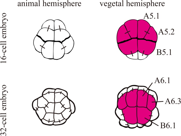
Fig 1. Expression of Foxd at the 16- and 32-cell stages.
Schematics of the animal and vegetal hemispheres of the bilaterally symmetrical 16-cell and 32-cell embryos. Cells expressing Foxd are colored in magenta. Their blastomere names are indicated in the right halves. Note that Foxd is expressed only at the 16-cell and 32-cell stages in early embryos. Black bars connecting two cells indicate their sister cell relationship.

