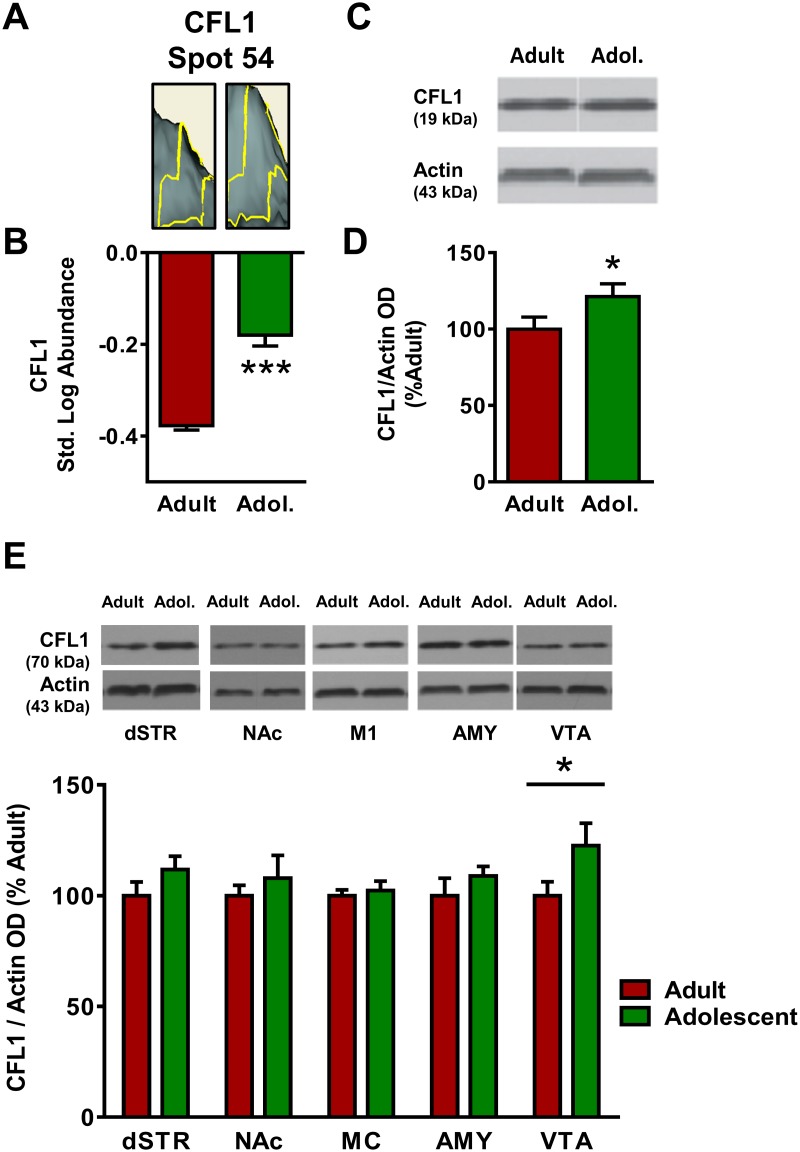
Fig 8. Adult and adolescent expression of cofilin-1 (CFL1).
(A) Representative 3D plot of CFL1 expression in adult (left) and adolescent (right) mice for Spot #54. (B) Standardized abundance (log) of CFL1 demonstrating higher expression in adults versus adolescents. (C) Representative gel image of a Western blot for CFL1 expression to confirm 2D-DIGE changes. (D) Quantification of Western blot results, confirming reduced expression of CFL1 (normalized to actin) in adolescents as compared to adults. (E) Top, representative gel images for each brain region; bottom, quantification of Western blots for each brain region. Adolescents show higher expression of CFL1 in VTA. There were no significant changes in CFL1 expression in dSTR, NAc, MC or Amy (p>0.05). (* indicates p≤0.05, *** indicates p≤0.001).

