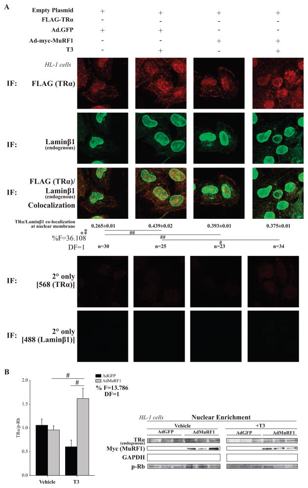
Figure 4.
MuRF1 inhibits T3-induced TRα localization to the nuclear envelope (lamin β1). HL-1 cardiomyocytes transfected with FLAG-TRα plasmid (empty FLAG = control), transduced with AdMuRF1 (AdGFP = control) for 4 h, and treated with 16.6 nM T3 for 2 h were assayed by confocal microscopy. (A) TRα (anti-FLAG, red) and endogenous lamin β1 (green) co-localization at the nuclear membrane. A two-way ANOVA test was used to determine statistical significance. *Significance on the level of adenovirus group, **Significance on the level of treatment group. The F statistic and degrees of freedom (DF) were reported when dependence between groups was found to be a significant source of variation. Significance between groups is represented as #P < 0.05, ##P < 0.001, as determined using a pairwise post-test. (B) HL-1 cardiomyocytes were transduced with AdMuRF1 (24 h) and treated with 16.6 nM T3 (2 h) followed by fractionation. Immunoblot of endogenous TR and myc-MuRF1 after T3 treatment (p-Rb used as a loading control) in nuclear fraction. Data represent three independent experiments. A two-way ANOVA test was used to determine statistical significance with a pairwise post-test. *Significance on the level of adenovirus group, % Significant interaction between adenovirus and treatment groups, #Difference from treatment control group (criterion for cutoff; P < 0.05). Data are presented as mean ± S.E.M., 400× final magnification.

