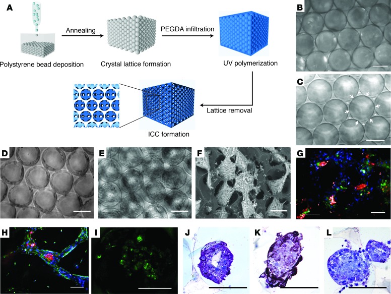
Figure 1. Fabrication of functional 3-dimensional hexagonally arrayed lobular human liver tissues.
(A) Schematic illustration of inverted colloidal crystal (ICC) scaffold fabrication. (B) Freestanding colloidal crystal lattice and (C) the resultant ICC with interconnected windows indicated by arrowheads and backbone of ICC indicated by asterisks. Morphology of fetal total liver cells in Col-I ICC (D) upon seeding and (E) 2 weeks after seeding. (F) Variable pressure scanning electron microscopy image demonstrating clusters of liver tissues with high interconnectivity across different ICC tiers. (G and H)Immunofluorescence imaging of engineered liver tissues 2 weeks after seeding for albumin (red) and DAPI (blue) costaining with (G) CK19 (green) or (H) vimentin (green). (I) Accumulation of Cholyl-L-lysyl-fluorescein (CLF) in liver tissues after 40 minutes of CLF incubation, followed by 40 minutes of washing. (J–L) Immunohistological images demonstrating heterogeneous populations within the engineered tissues stained for albumin (J), CK19 (K), and CD68 (L). Scale bars: 100 μm.

