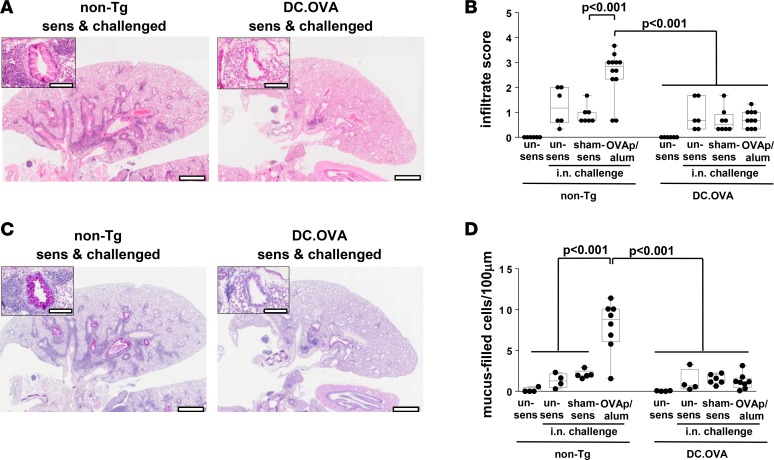
Figure 3. Expression of allergen in DC prevents pathology.
DC.OVA or nontransgenic littermates (non-Tg) were sensitized with OVA323-339/alum (OVAp/alum; d0, d14), were sham-sensitized with PBS/alum (sham-sens), or were left unsensitized (un-sens) and i.n. challenged with OVA (days 11–14 and 19–24 after sensitization). One day after the last i.n. challenge, mice were euthanized for analysis. Lungs were collected and H&E (A) or periodic acid–Schiff (PAS) (C) stained, and images were analyzed to define cellular infiltrate (B) and mucus-filled goblet cell frequency (cells per 100 μm basement membrane) (D). Scale bars (A and C) depict 1 mm (low-power) and 200 μm (high-power inset). Data are representative micrographs from 4–6 mice per group pooled from 3 experiments or show values for individual mice pooled from 3 experiments, with box and whisker plots showing median, quartiles, and range. ANOVA/Tukey’s post-test

