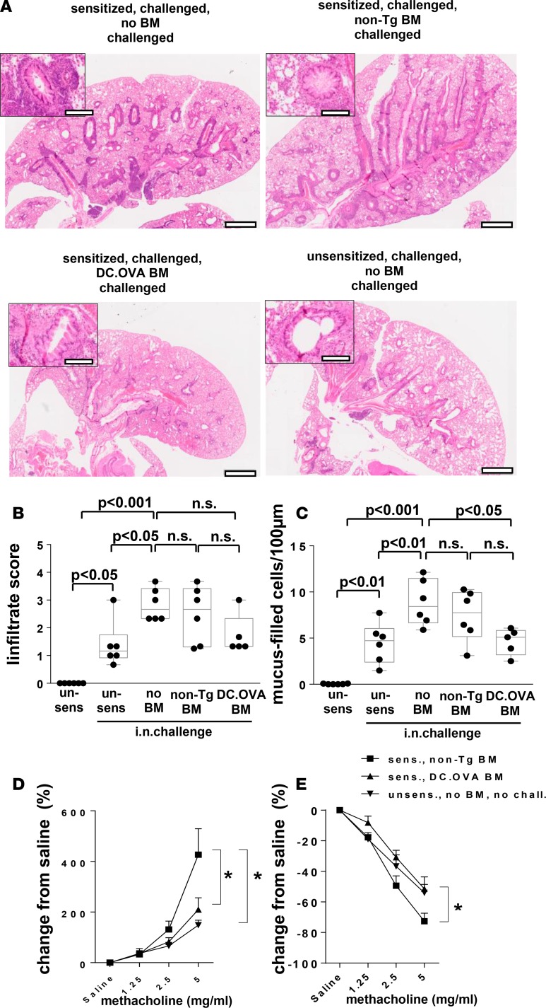
Figure 7. Transfer of OVA-encoding BM ameliorates established airway inflammation.
(A–E) BALB/c mice were sensitized twice with OVA323-339/alum (days 0, 14) or not (un-sens) and challenged with OVA i.n. daily (days 11–14 and 19–24 after sensitization). One week later, some mice were irradiated (300 cGy) and injected with BM from DC.OVA (DC.OVA BM) or non-Tg (non-Tg BM) donors. Four weeks later, mice were i.n. challenged with OVA (5 days i.n., 4 days rest, 5 days i.n.). One day after the last i.n. challenge, mice were euthanized for analysis. (A–C) Untreated BALB/c mice (un-sens) were included for comparison. Lungs were collected and H&E or periodic acid–Schiff (PAS) stained (A), and images were analyzed to define cellular infiltrate (B) and mucus-filled goblet cell frequency (cells/100 μm basement membrane) (C). Scale bars (A) depict 1 mm (low-power) and 200 μm (high-power inset). (D and E) Mice were intubated, and changes in airways resistance and compliance in response to challenge with increasing doses of methacholine were measured. Data are representative micrographs of 6 mice per group from 2 experiments (A) or show values for individual mice pooled from 2 experiments, with box and whisker plots showing median, quartiles, and range (B and C) or mean ± SD for 8 mice per group pooled from 2 experiments (D and E). ANOVA/Newman-Keuls post-test. *P < 0.05.

