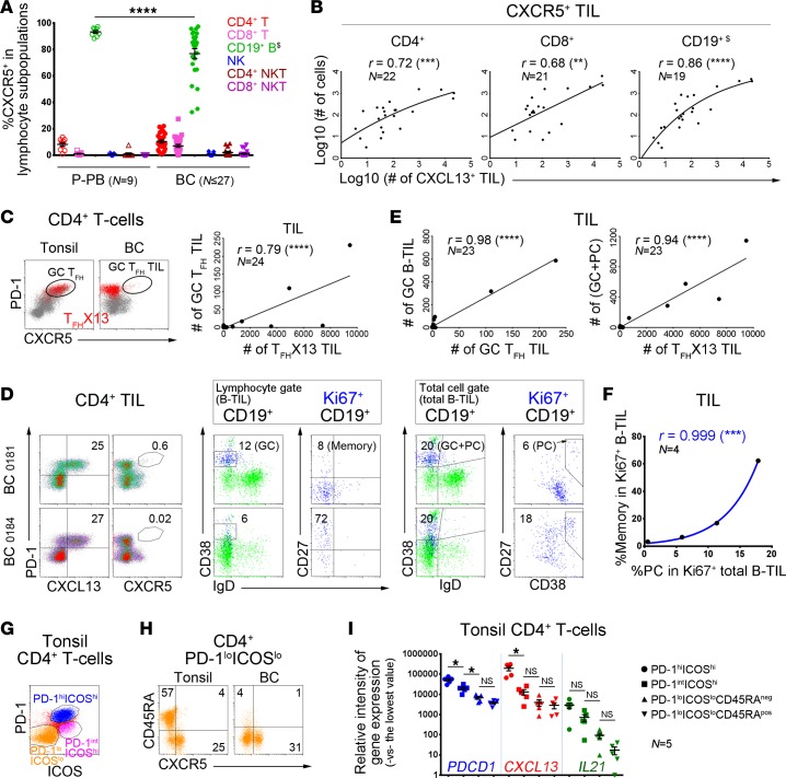
Figure 6. TFHX13 tumor-infiltrating lymphocytes (TIL) in human breast cancer (BC) are correlated with specific B-TIL maturation stages.
(A–H) Flow cytometric analyses. (A) CXCR5 expression in specific lymphocyte subpopulations from patient peripheral blood (P-PB) and BC (n = range 16–27 BC). One-way ANOVA followed by Tukey’s test. (A–D) Gating strategies are shown in Supplemental Figure 6A; $B cells were analyzed in the total cell gate. (B) Correlation between the number of CXCR5+ cells in specific TIL subpopulations and total CXCL13+ TIL in fresh BC homogenates. (C) Germinal center (GC) TFH (PD-1hiCXCR5hi) or TFHX13 (CXCL13+CD4+; red) cells in tonsils and BC (dot plots); correlation between #GC TFH and #TFHX13 BC TIL (graph). (D) Equally high TFHX13 TIL responses (left) but distinct proliferating (Ki67+) B-TIL subpopulations (middle and right) in 2 BC. B cell differentiation markers are shown for total CD19+ (green) and Ki67+CD19+ (blue) B-TIL (GC B cells = CD38+IgD–; plasmablasts/plasma cells [PC] = CD38hiIgD– or CD38hiCD27int/hi; CD27 = memory B cell marker). (E) Correlation between #GC TFH and #TFHX13 TIL defined in C and #B-TIL subpopulations defined in D. (F) Correlation between % memory cells (excluding PC) in Ki67+ B-TIL (lymphocyte gate) and PC in Ki67+ total B-TIL (total cell gate) from 4 BC with sufficient numbers of Ki67+ B-TIL (gating strategies in Supplemental Figure 6B). (G) PD-1/ICOS–defined CD4+ T cell subpopulations from tonsils. (H) CD45RA and CXCR5 expression on PD-1loICOSloCD4+ T cells from tonsils and BC. (I) Expression of PDCD1 (PD-1), CXCL13, and IL21 genes (qPCR) in CD4+ T cell subpopulations sorted from 5 tonsils. One-way ANOVA followed by Sidak’s test. *P < 0.05, **P < 0.01, ***P < 0.001, ****P < 0.0001.

