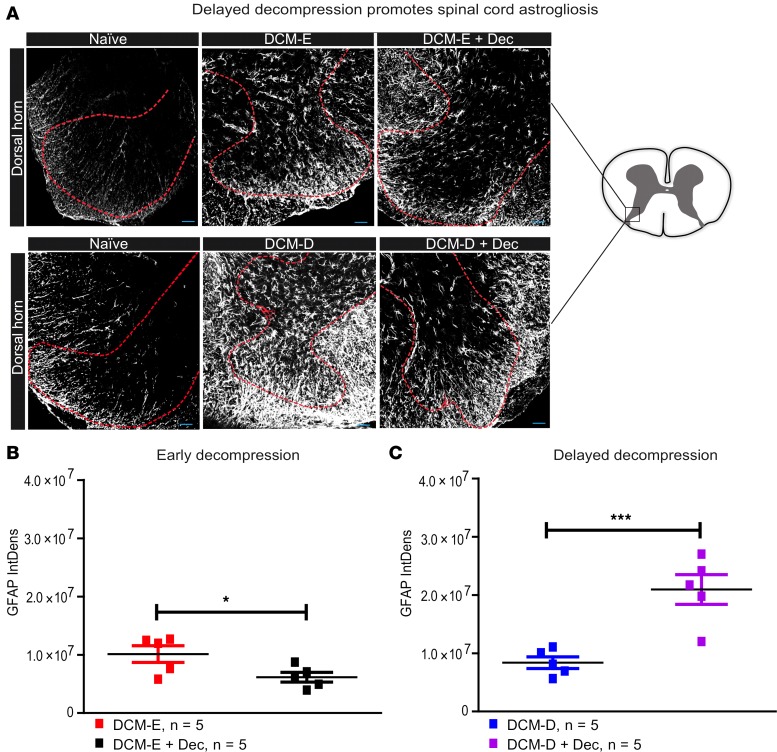
Figure 8. Early decompression attenuates astrogliosis in the dorsal horns.
(A) Representative confocal images of dorsal horns from mice that underwent early or delayed decompression, their age-matched sham controls, and age-matched naive mice, all stained for glial fibrillary acidic protein (GFAP). The red dotted lines delineate the dorsal horns where GFAP immunoreactivity was quantified. The spinal cord diagram on the right side represents the area of constant size within the dorsal horns, used for the analysis of GFAP immunoreactivity. (B) GFAP immunoreactivity was significantly reduced at 5 weeks after decompression in the DCM-E + Dec (n = 5) group compared with the age-matched DCM-E group (n = 5). *P < 0.05, Mann-Whitney U test. (C) Astrogliosis was significantly increased in the dorsal horns of DCM-D + Dec (n = 5) compared with the DCM-D group (n = 5). ***P < 0.001, Mann-Whitney U test. All the results are presented as mean ± SEM. Scale bars: 25 μm. DCM, degenerative cervical myelopathy; Dec, decompression; DCM-E, age-matched early sham decompressed group; DCM-D, age-matched delayed sham decompressed group; IntDens, integrated density.

