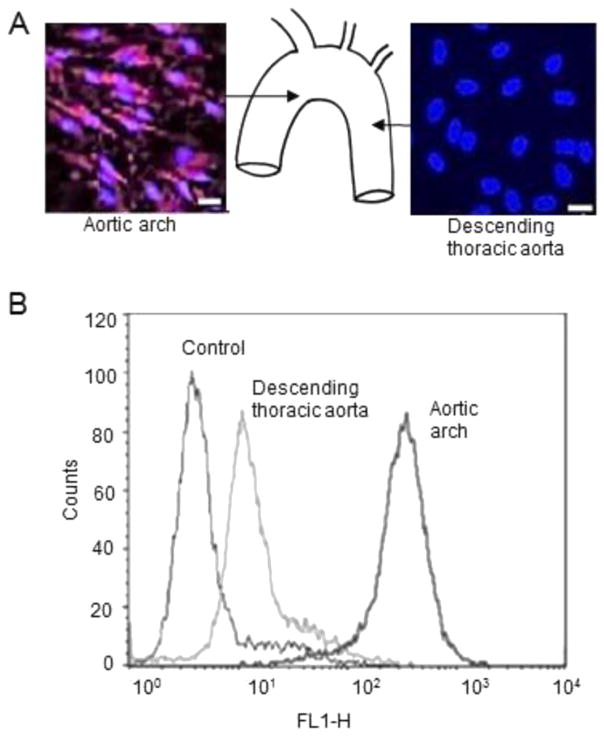
Fig. 1.
Differential expression of VCAM-1 in regions of the mouse aorta. A, representative en face immunostaining for VCAM-1 expression in the aorta of wild-type mice. Endothelial cell nuclei and VCAM-1 are in blue and green, respectively. Scale bar, 20 μm. P < 0.05. n = 15 visual fields. B, Flow cytometry analysis of EC surface expression of VCAM-1 in the aortic sinus and descending thoracic aorta of wild-type mice. Graphs show that the number of cells expressing VCAM-1 on the cell surface was greater in the aortic sinus as compared to the descending aorta. Endothelial cells incubated with only a second antibody were used as controls.

