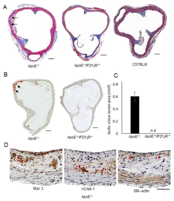
Fig. 5.
Analysis of atherosclerotic lesions in the aortic sinus of male ApoE−/− and ApoE−/−/P2Y2R−/− littermate mice fed standard chow diet for 15 weeks. A, Aortic sinus cross sections stained with Masson’s trichrome for gross morphological analysis and classification of lesions as shown in Table 2. B–C, Cross sections were stained with Oil Red O and the relative lesion area was calculated by dividing the lesion area by the total cross-sectional area. D, Representative images of immunohistological staining of atherosclerotic lesions in the aortic sinus. Adjacent sections were stained with, Mac-3 antibody, VCAM-1 and smooth muscle α-actin antibodies, respectively. Each scale bar represents 100 μm.

