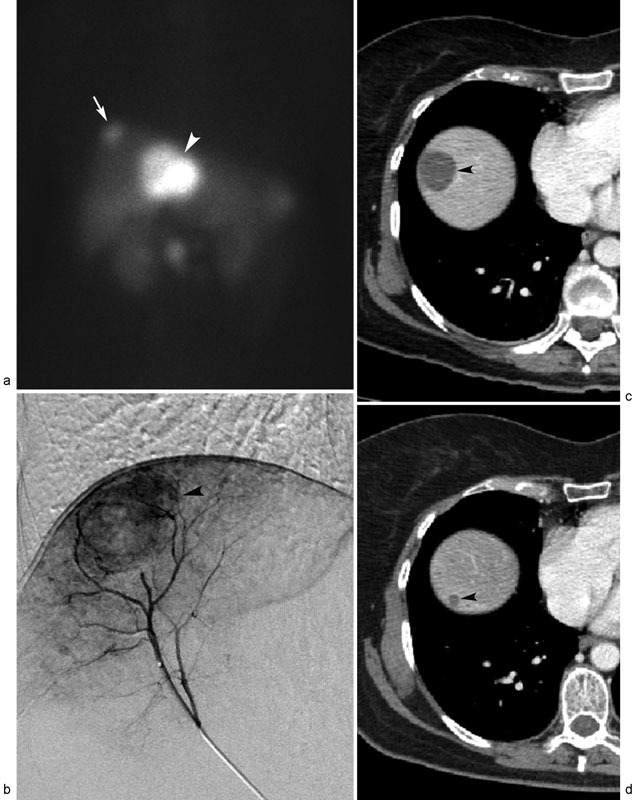
Fig. 2.

Metastatic neuroendocrine tumor treated with transarterial chemoembolization (TACE). Octreotide scan ( a ) displays oligonodular liver metastases spanning segment 4 (arrowhead) and segment 8 (arrow) tumors. ( b ) Digital subtraction arteriogram performed from segment 8 hepatic artery reveals hypervascular dome tumor (arrowhead), which was treated with c-TACE. Successive posttreatment contrast-enhanced CT scans performed 1 week ( c ) and 1 year ( d ) after TACE demonstrate gradual, near-complete regression of necrotic right hepatic lobe liver metastasis (arrowheads).
