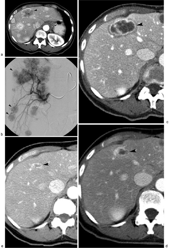
Fig. 3.

Metastatic neuroendocrine tumor treated with 90 Y RE. Contrast-enhanced CT scan ( a ) shows multifocal liver metastases, with index tumor (arrowhead) located in liver segment 4. Digital subtraction right hepatic arteriography ( b ) displays multifocal hypervascular right hepatic lobe liver metastases (arrowheads), which were treated with 90 Y-labeled resin microspheres. Sequential posttreatment contrast-enhanced CT scans performed 3 months ( c ), 6 months ( d ), and 1 year ( e ) after 90 Y RE demonstrate progressive, near-complete regression of necrotic segment 4 liver metastasis (arrowheads).
