
Figure 1.
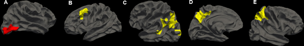
Cortical thickness associations between (A) BP and right lateral occipital; (B) Chol and left caudal medial frontal; (C) MetabDys and left inferior parietal; (D) MetabDys and left precuneus; (E) MetabDys and right superior parietal NOTE: Whole brain cortical thickness results derived from FreeSurfer. Maps were smoothed with a 15mm full width half maximum Gaussian kernel and correction for multiple comparisons was performed using the Monte Carlo Null-z simulation (p<0.05). All analyses have been adjusted for age and sex. Red indicates negative associations with cortical thickness and yellow indicates positive associations with cortical thickness.