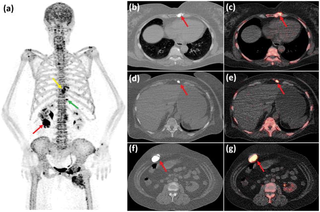
Figure 1.
A 67-year-old woman with established diagnosis of melorheostosis since the age of 22 was referred to our institute. Although independent in self-care activities, the patient has been suffering from chronic pain secondary to the melorheostosis with three to five episodes of pain flare-ups per year. The patient developed lymphedema of the left lower extremity and leg length discrepancy with the left leg being longer. In order to assess the disease activity the patient underwent whole body 18F-NaF PET/CT scan, which showed multiple 18F-NaF avid lesions corresponding to the hyperdense bone abnormalities and the soft-tissue ossifications. A prominent lesion with intensely elevated 18F-NaF activity (SUVmax: 45,4) resided in the left lower sternum (Fig. 1a: Maximum Intensity Projection (MIP) PET image of the torso, yellow arrow; Fig 1b & 1c: axial CT and axial fused PET/CT images of the chest, red arrows). Also, two sites of extra-osseous bone formation were observed in the anterior abdominal wall (Fig. 1a: green arrow; Fig 1d & 1e: axial CT and axial fused PET/CT images of the upper abdomen, red arrows) and in the right anterolateral abdominal wall (Fig. 1a: red arrow; Fig 1f & 1g: axial CT and axial fused PET/CT images of the abdomen, red arrows) which also showed markedly elevated 18F-NaFuptake (SUVmax: 30,3 & 42,6 respectively).

