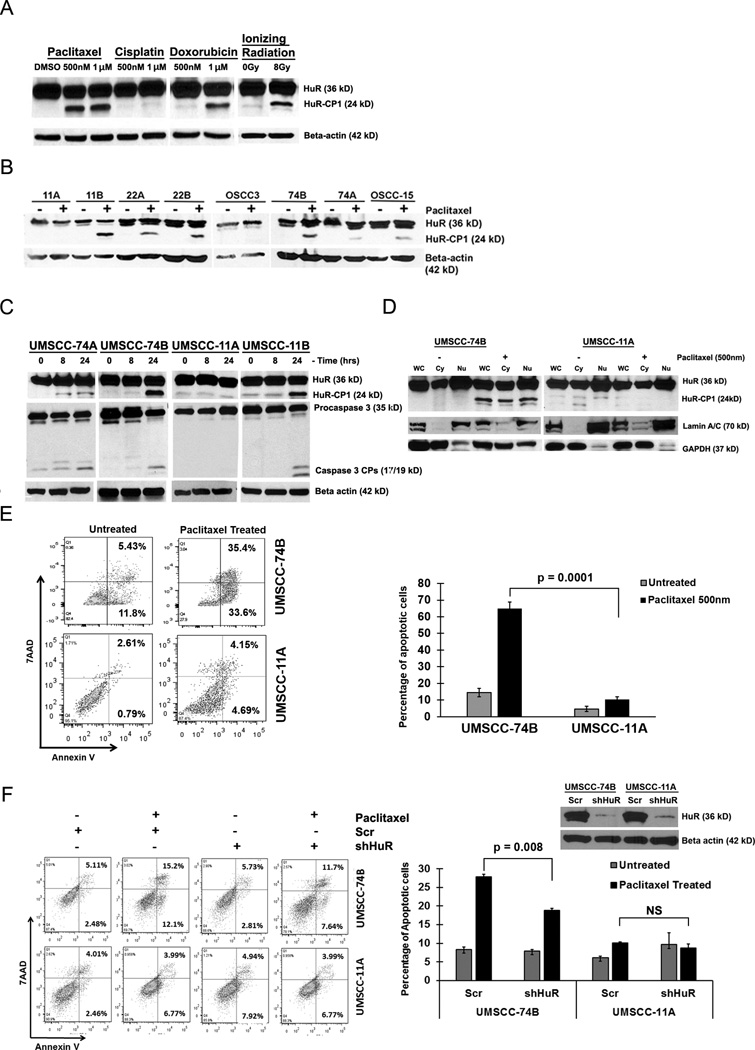
Figure 1. Caspase-3 mediated cleavage of HuR is cancer cell specific.
A) Western blot analysis of HuR cleavage patterns in UMSCC-74B cells treated with either DMSO, paclitaxel (0.5µM-1µM), cisplatin (1µM), doxorubicin (1µM) or 8Gy of ionizing radiation. B) Western blot analysis of HuR in multiple oral cancer cells treated with DMSO and paclitaxel (0.5µM). C) Western blot analysis of HuR and caspase-3 oral cancer cell lines treated with 0.5µM paclitaxel at the indicated time points. β-actin: loading control. D) Western blot analysis of HuR expression in nuclear/cytoplasmic fractionation during paclitaxel treatment. Lamin A/C (nuclear marker) and GAPDH (cytoplasmic marker). Representative western blots from three independent experiments are shown. E) Apoptotic rate of 74B and 11A cells treated with DMSO or 0.5µM paclitaxel for 24hrs. The percentage of early (bottom right quadrant) and late (top right quadrant) apoptotic cells are depicted in the scatter plots and bar graphs. Data represented as mean ± SD; N=3. F) Apoptotic rate between HuR silenced 74B and 11A cells under DMSO or 0.5µM paclitaxel treatment for 16hrs measured by flow cytometry. Data represented as mean ± SD; N=3.

