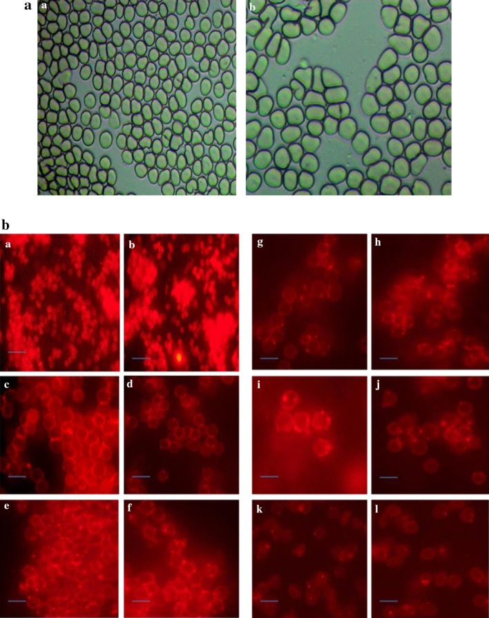
Fig. 3.
a Morphological changes in human erythrocytes treated with magnetosomes (a) Human erythrocytes treated with PBS as control (b) Human erythrocytes treated with magnetosomes (150 µg/ml). b Effect of different concentrations of magnetosomes on human erythrocytes morphology. Microscopic images of RBC’s (a and b) erythrocytes treated with PBS as control (c and d) RBC’s treated with 10 µg/ml magnetosomes, (e and f) RBC’s treated with 20 µg/ml magnetosomes, (g and h) RBC’s treated with 50 µg/ml magnetosomes, (i and j) RBC’s treated with 100 µg/ml magnetosomes, (k and l) RBC’s treated with 150 µg/ml magnetosomes. (Magnification: ×40)

