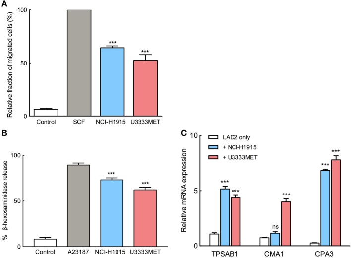
Figure 2.
Migration and activation of mast cells (MCs) in response to brain metastasis (BM) cells. (A) Migration of MCs toward NCI-H1915 or U3333MET cells measured by transwell assay. Medium without serum was used as negative and SCF as positive chemotactic control. (B) β-hexosaminidase release by MCs in response to stimulation by NCI-H1915 or U3333MET cell lines as indicated. 2 µM calcium ionophore A23187 was used as a positive control and HBSS as negative control. (C) Quantitative PCR to evaluate mRNA levels of the MC-specific proteases in response to the indicated BM cell line. Unstimulated LAD2 cells were used as control. β-actin mRNA detection was used for normalization. The experiments were performed three times in triplicates and mean values + SEM was plotted, ns, not significant; **p < 0.01, ***p < 0.001.

