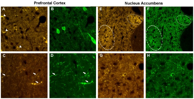
Figure 4.
Phenotype of the VGF positive neurons. Prefrontal cortex. Using the VGF C-terminus (A) and the parvoalbumin (B) antibodies, an incomplete colocalization profile was found in the cell bodies, with some perikarya labeled by the VGF C-terminus antibody only (identified by the arrows, A). Almost all the TLQP positive nerve axons (C) contained also tyrosine hydroxylase (TH; D, identified by the arrows). Nucleus accumbens. C-terminus-immunoreactivity (E) was revealed in a subpopulation of neuron terminals containing glutamic acid decarboxylase (GAD; F) inside specific areas of the shell (identified by the circles). The N-terminus immunoreactivity (G) was almost completely found in all axons-, perikarya- and nerve terminals-secreting somatostatin (H). VGF antibody: Cy3 red labeling, parvoalbumin, somatostatin, GAD: Cy2 green labeling. Magnification: 600× (A–D,G,H) and 400× (E,F).

