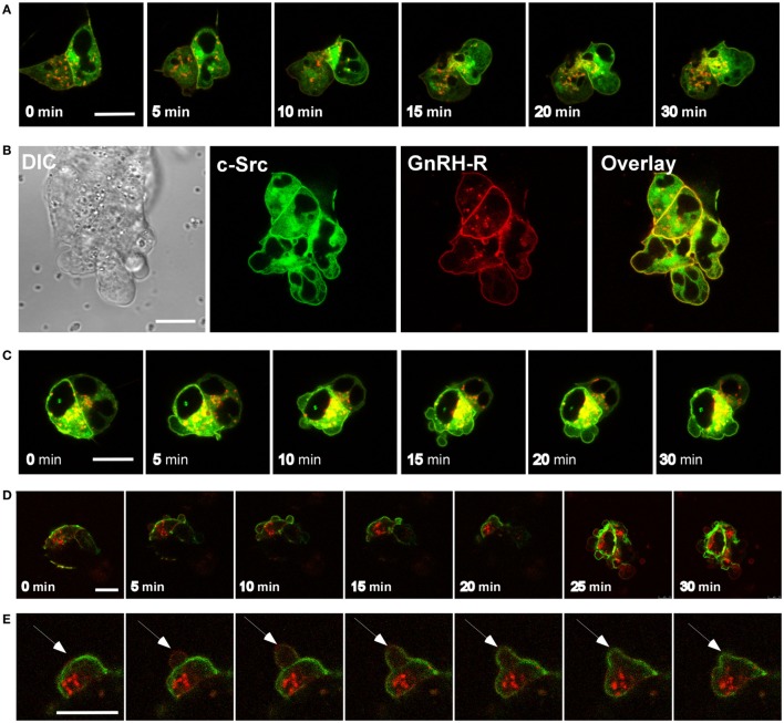
Figure 5.
c-Src and vinculin are present in the blebs. (A) Addition of GnRH (0–30 min, 10 nM) to serum-starved LβT2 cells transfected with c-Src-GFP and GnRH receptor (GnRHR)-mCherry resulted in bleb formation, while c-Src is present in the blebs. The scale bar is 10 µm. (B) Blebs time laps of DIC and fluorescent images of Src-GFP, GnRHR-mCherry, and overlay showing colocalization of c-Src and the GnRHR in the blebs. The scale bar is 10 µm. (C) Serum-starved LβT2 cells transfected with c-Src-GFP and GnRHR-mCherry were pretreated with the c-Src inhibitor PP2 (10 µM) for 30 min. Thereafter, GnRH (10 nM) was added for 30 min. Similar results were observed in two other experiments. The scale bar is 10 µm. (D) Addition of GnRH (0–30 min, 10 nM) to serum-starved LβT2 cells transfected with GnRHR-mCherry and vinculin-GFP resulted in bleb formation, while vinculin is present in the blebs. The scale bar is 10 µm. (E) Unlike c-Src, ERK1/2, focal adhesion kinase, and paxillin (see below), vinculin was recruited to the blebs during stabilization and retraction. Addition of GnRH (10 nM) to serum-starved LβT2 cells transfected with GnRHR-mCherry and vinculin-GFP resulted in bleb formation, single blebs were monitored every 10 seconds. The scale bar is 10 µm.

