Figure 7.
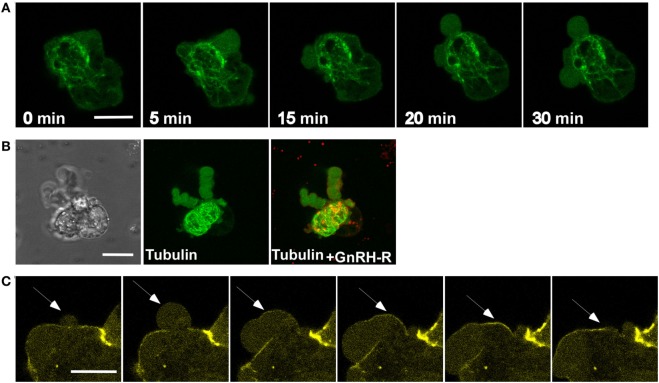
Tubulin and actin, but not microtubules, are present in the blebs. (A) Images from a confocal microscopy time-lapse movie of serum-starved LβT2 cells transfected with GnRH receptor (GnRHR)-mCherry and EMTB-3XGFP (the microtubule-binding domain of ensconsin (EMTB) fused to three GFP molecules, allowing microtubules visualization) and treated with GnRH (30 min, 10 nM). Bleb formation was noticed, while microtubules are not present in the blebs. The scale bar is 10 µm. (B) Blebs images after GnRH treatment, including DIC and fluorescent images of GnRHR-mCherry and EMTB-3XGFP, supporting the data observed in panel (A). The scale bar is 10 µm. (C) Actin is involved in bleb retraction. Addition of GnRH (30 min, 10 nM) to serum-starved LβT2 cells transfected with actin-YFP resulted in bleb formation. Actin is recruited to the blebs after they are stabilized and is best observed during blebs retraction (see arrows). The scale bar is 10 µm.

