Figure 6.
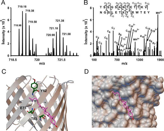
Photolabeling a third residue, Tyr62, in the cholesterol binding pocket in E73Q mVDAC1. A, an MS1 doublet corresponding to the peptide TSSGSANTETTKVNGSLETKYRWTEY from E73Q mVDAC1 photolabeled with KK174 (z = 5) and coupled to light and heavy FLI-tag. B, fragmentation (ETD) spectra corresponding to the feature in A. Site defining ions localize KK174 photolabeling to Tyr62. C, the same cholesterol binding pose as in Fig. 2E showing Tyr62, Glu73, and Thr83. D, space filling view of the binding pocket mapped by KK174 and LKM38.

