Figure 7.
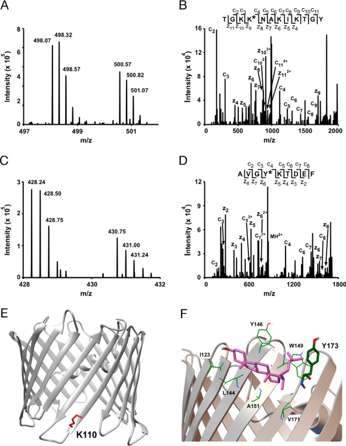
Additional sites of KK174 photolabeling in E73Q mVDAC1. A, an MS1 doublet corresponding to the peptide TGKKNAKIKTGY labeled with KK174 (z = 4) and coupled to light and heavy FLI-tag. B, fragmentation (ETD) spectra corresponding to the heavy feature in A. Site defining ions localize KK174 labeling to Lys110. C, an MS1 doublet corresponding to the peptide AVGYKTDEF labeled with KK174 (z = 4) and coupled to light and heavy FLI-tag. D, fragmentation (ETD) spectra corresponding to the light feature in C. Site defining ions localize KK174 labeling to Tyr173. E, mVDAC1 crystal structure (PDB code 3emn) highlighting the photolabeled residue Lys110. F, a cholesterol binding pose from Autodock located within a previously identified cholesterol binding pocket (8) formed by Val171, Ala151, Leu144, Ile123, Tyr146, and Trp149. The aliphatic tail of cholesterol in this pose is near Tyr173, consistent with KK174 labeling of this residue in E73Q mVDAC1.

