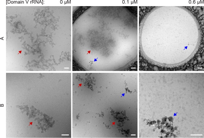
Figure 6.
Cryo-TEM images of p53C aggregates. The p53C samples (4.5 μm) alone or with different concentrations of dom V rRNA were incubated for 120 min at 37 °C and then subjected to cryo-TEM. Blue arrows indicate ice crystals, whereas red arrows indicate p53C aggregates. Row A includes the global view of large aggregates, whereas row B is composed of images of smaller-sized aggregates. In the case of 0.6 μm dom V rRNA, no aggregates were detected. The scale bar corresponds to 100 nm.

