Figure 1.
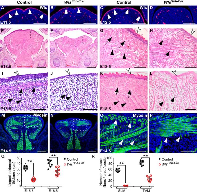
Epithelial Wnt production regulates embryonic tongue development. A–D, immunohistochemical staining analysis for Wls expression in the dorsal epithelium (arrowheads) and mesenchyme of embryonic tongues at E11.5 and E12.5. E–L, histological analyses of embryonic tongues show that deletion of epithelial Wls leads to microglossia (E and F), epithelial hypoplasia (white arrows), and compromised muscle formation. SLM and transverse and vertical myofibers (TVM) are indicated by arrows and arrowheads, respectively. Boxed regions in E and F are shown magnified in G and H. E–H, frontal sections; I–L, sagittal sections. M–P, immunostaining of myosin (green) for muscle fibers in E14.5 tongues. Boxed regions in M and N are shown magnified in O and P. Q, statistical analysis of lingual epidermal thickness from histological sections (I–L). R, comparison of the number of SLM and transverse and vertical myofibers in the designated area (O and P) of control and Wls mutant tongues. Data are shown as scatter plots. **, p < 0.01. Scale bars, 200 μm (A–D, M, and N), 500 μm (E and F), and 100 μm (G–L, O, and P).

