Figure 2.
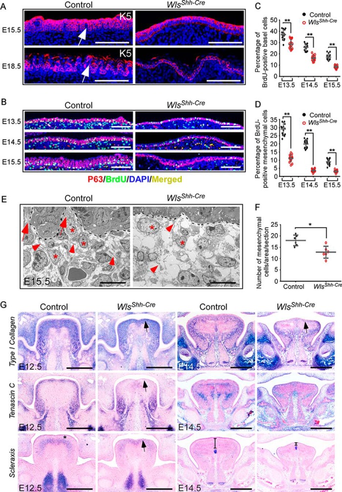
Loss of epithelial Wls impairs the epithelial basal cell proliferation and the lamina propria formation in epithelial Wls mutant tongues. A, immunofluorescence (red) of K5 expression for basal cells in dorsal lingual epithelium at E15.5 and E18.5. Prominent lamina propria cores are indicated by arrows. B, immunostaining of BrdU (green) and p63 (red) shows BrdU-labeled cells in basal cells from E13.5 to E15.5. C and D, percentage of BrdU-labeled cells in p63-positive basal cells (C) and underlying tissue (D) in a designated area of the tongue (B). E, TEM micrographs show connective tissue cells (*) and fibers (arrowheads) in E15.5 control and WlsShh-Cre tongues. Lamina propria cores are indicated by arrows. F, quantitation of connective tissue cells from TEM micrographs of E15.5 control and Wls mutant tongues. G, in situ hybridization for the type I collagen, tenascin C, and scleraxis transcripts at E12.5 and E14.5. Data are shown as scatter plots. *, p < 0.05; **, p < 0.01. Scale bars, 100 μm (A and B), 10 μm (E), and 400 μm (G).

