Figure 3.
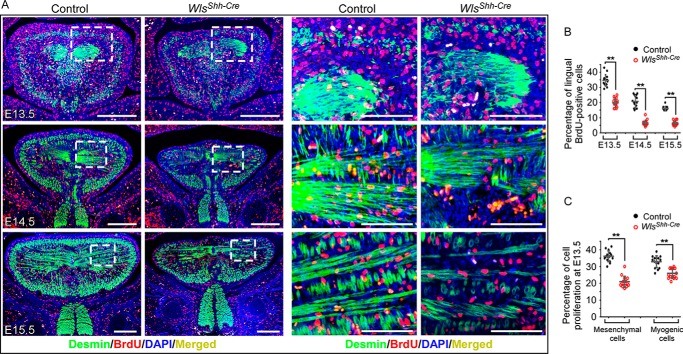
Epithelial Wnt production mediated by Wls regulates cell proliferation in embryonic tongues. A, immunofluorescence with antibodies against desmin (green) and BrdU (red) shows BrdU-labeled cells in muscle (desmin-positive) and the surrounding tissue. Boxed areas are enlarged on the right. B, percentage of BrdU-positive nuclei in the total cell population in tongues from E13.5 to E15.5. C, percentage of BrdU-positive nuclei in desmin-positive cells (myogenic cells) and desmin-negative mesenchyme cell in E13.5 tongues. Data are shown as scatter plots. **, p < 0.01. Scale bars, 100 μm (enlarged images of boxed regions in A); 250 μm (others).

