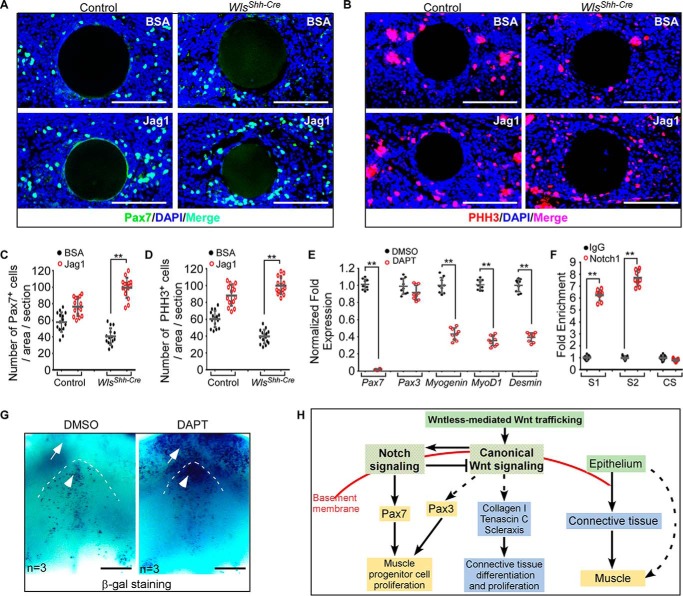
Figure 6.
Reciprocal interaction of Wnt and Notch signaling regulates embryonic tongue development. A and B, immunostaining analysis for Pax7 (A) and pHH3 (B) expression in the tongue explants implanted with Jag1 or BSA-soaked beads. C and D, quantitation of the number of Pax7-positive (C) and pHH3-positive (D) cells in the tongue explants. E, quantitative analysis of the myogenic gene expression in tongue explants after incubation with or without DAPT. F, -fold enrichment of RBP-J-binding sites (S1 and S2) and control site (CS) of the Pax7 promoter region following ChIP with a Notch1 antibody. G, BATGAL staining for canonical Wnt activity in tongue explant cultured with or without DAPT. H, schematic showing a genetic hierarchy that regulates embryonic tongue development. Data are shown as scatter plots. **, p < 0.01. Scale bars, 100 μm (A and B) and 200 μm (G).

