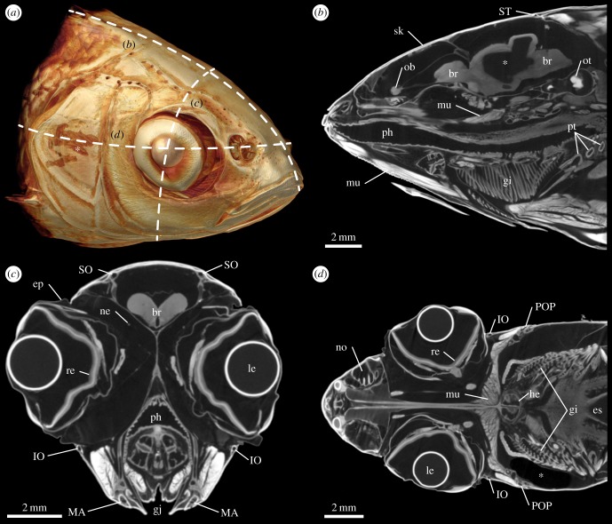
Figure 2.
External and internal anatomy of the head of Leuciscus idus analysed using contrast-enhanced µCT. (a) Anterolateral view of a volume rendering illustrating the position of (b) sagittal, (c) transverse and (d) coronal virtual sections. Asterisk in (a) indicates a lesion of the skin caused during specimen handling. Asterisk in (b) depicts an area inside the brain with insufficient staining. Asterisk in (d) indicates a region with low X-ray absorption due to the presence of an air bubble. br, brain; ep, epithelium; es, oesophagus; gi, gill; gj, gill juncture; he, heart; IO, infraorbital canal; le, lens; MA, mandibular canal; mu, muscle tissue; ne, nerve tissue; no, nostril; ob, olfactory bulb; ot, otolith; ph, pharynx; POP, preopercular canal; pt, pharyngeal tooth; re, retina; sk, skeleton; SO, supraorbital canal; ST, supratemporal commissure.

