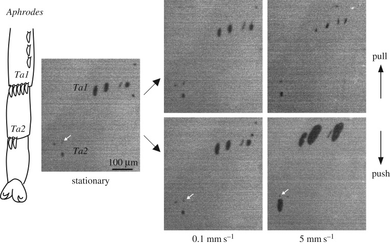
Figure 5.
Contact area of hind leg platellae of Aphrodes during shear force measurements at different sliding velocities. Platellae were brought into contact with a normal force of 5 mN (‘stationary’) and then sheared in the pushing and pulling direction for 2 s, while keeping the normal force constant. Images show contact areas 0.2 s before the end of the sliding movement. Ta1: tarsomere 1, Ta2: tarsomere 2. White arrows show the tip of the tarsal spine adjacent to the platella on Ta2. It can be seen that the contact area of the platella expanded both laterally and longitudinally by elongating mainly on the proximal side. The scale bar applies to all images.

