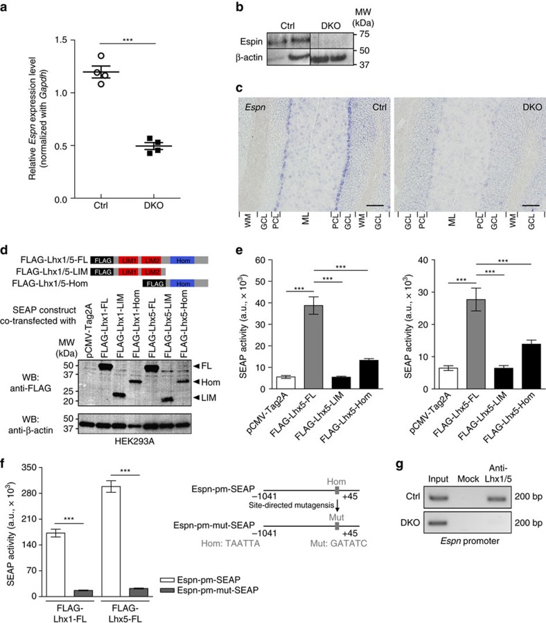
Figure 3. Lhx1/5 transcriptionally regulate Espn.
(a) Real-time quantitative PCR showing the relative Espn expression level in the adult DKO mutant cerebellum when compared with the control cerebellum; n=4 mice per group; t-test; ***P<0.001. (b) Western blots showing the cerebella of the DKO mutants had lower Espin level than the controls. The experiment was repeated twice, seven mice for each genotype. (c) In situ hybridization for Espn transcripts showing the PCs of the DKO mutants (right) had a lower Espn expression than the controls (left). The experiment was repeated three times with three mice per genotype. ML, molecular layer; PCL, Purkinje cell layer; GCL, granule cell layer; WM, white matter. Scale bars, 100 μm. (d) Schematic illustrations showing different forms of FLAG-tagged Lhx1/5 proteins. Western blots showing the coexpression of Lhx1/5 proteins with SEAP. FL, full-length; LIM, LIM domains; Hom, homeodomain. (e) Bar graphs showing the differences in SEAP activity under the control of the Espn promoter (Espn-pm-SEAP) when different forms of Lhx1 (left) and Lhx5 (right) proteins were coexpressed. Analysis of variance (ANOVA); ***P<0.001. (f) Schematic illustrations showing the SEAP plasmid containing an Espn promoter with mutation (Mut) in the binding site of Lhx1/5 homeodomain (Hom). Bar graphs reveal the differences in the SEAP activity with and without the mutation in the Espn promoter. For all SEAP assays, at least three independent replications were performed and n=9 for each group of experiments; t-test; ***P<0.001. (g) Chromatin immunoprecipitation (ChIP)-PCR showing the in vivo binding of Lhx1/5 on Espn promoter in the controls (top) but not in the DKO mutants (bottom); n=2 mouse cerebella pooled for each group. The ChIP experiment was repeated three times, each with two different mouse cerebella per group.

