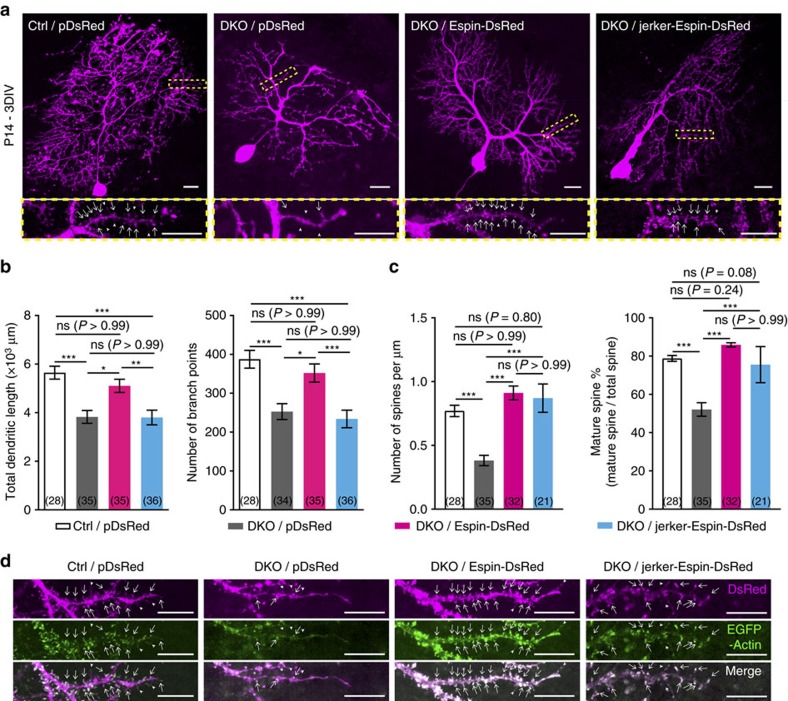
Figure 8. Overexpression of Espin rescues the dendritic defects in the PCs of the DKO mutants ex vivo.
(a) Representative PCs from P14 cerebellum slice cultures of the DKO mutants transfected with pDsRed or Espin-DsRed or jerker-Espin-DsRed and collected at 3 days in vitro (DIV). PCs from P14 control mice transfected with pDsRed served as the controls. Zoom-in images of yellow boxed regions show the dendritic spines of the respective PCs. Note that the dendritic trees of the mutant PCs transfected with Espin-DsRed were more extensive and had more mature spines. Scale bars, 20 μm; 10 μm (zoom-in). (b) Bar graphs showing the total dendritic length (left) and the branching number (right) of transfected PCs. The dendrite elongation and branching of the mutant PCs restored to the controls' level after transfection of Espin-DsRed. (c) Bar graphs showing the density of dendritic spines (left) and the percentage of mature spines (right). Spinogenesis and spine maturation of the mutant PCs significantly increased after transfection of Espin-DsRed or jerker-Espin-DsRed. Brackets show the number of transfected PCs analysed. N=4–9 mice. Kruskal–Wallis test; ns, not significant; *P<0.05, **P<0.01 and ***P<0.001. (d) Distal dendrites of PCs from the controls or the mutants co-transfected with EGFP-Actin and pDsRed or Espin-DsRed or jerker-Espin-DsRed. White arrows point to the mature spines while arrowheads point to the immature spines. Both normal Espin and jerker-Espin were highly colocalized with F-actin in the dendritic spines of the transfected PCs. Scale bars, 10 μm.

