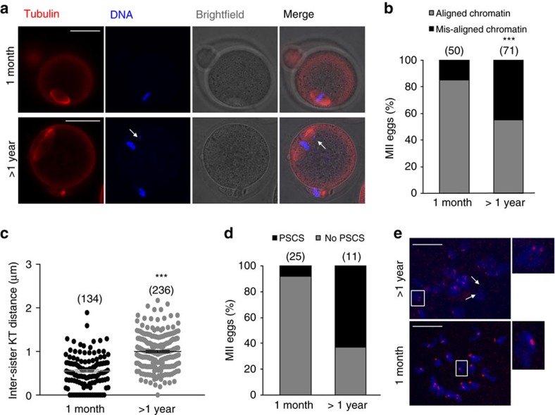
Figure 1. Chromosome misalignment and reduced sister chromatid cohesion in MII eggs from aged MF1 mice.
Representative images of immunostaining for DNA in blue and tubulin in red (a), and quantification of chromosome misalignment (b) in MII eggs from young (1 month) and aged (>1 year) mice. The number of eggs used is shown in parentheses. The arrow points towards misaligned DNA. (c) The mean distance between sister kinetochores in MII eggs from 1 month (n=14 eggs) and >1 year (n=20 eggs) old mice. The number of kinetochores measured is shown in parentheses. In b,c, the results are mean±s.e.m. ***P<0.001. P values were calculated with one-sided Student's t-test. (d) Rates of PSCS in MII eggs from 1 month- versus >1 year old mice. The number of eggs used is shown in parentheses. (e) Representative example of the chromosome spreads assay demonstrating MII eggs from >1 year- (top panel) and 1 month old (bottom panel) mice. DNA is shown in blue and CREST-labelled kinetochores are shown in red. The arrows point towards two-separated sister chromatids, and the insets show the difference in the inter-sister kinetochore distance between eggs from 1 month- and >1 year old mice. Scale bars, 50 μm. Results are from three to four independent experiments involving two to five mice per experimental group.

