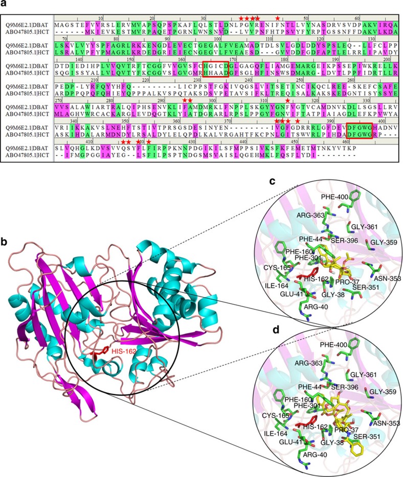
Figure 4. The DBAT structure predicted by homology modeling (template: HCT) and molecular docking of DBAT with the substrates.
(a) Sequence alignment between DBAT and the template HCT (ABO47805.1). Identical and similar residues are highlighted in green and pink, respectively. Residues predicted to be involved in acyl accepter substrate binding are highlighted with red stars, and the conserved HXXXD and DFGWG motifs are marked in red box. (b) Homology model of DBAT constructed with Swiss-Model (the distance 5 Å around the substrate position was circled). (c) Enlarged view of molecular docking of DBAT with 10-DAB. (d) Enlarged view of molecular docking of DBAT with DT.

