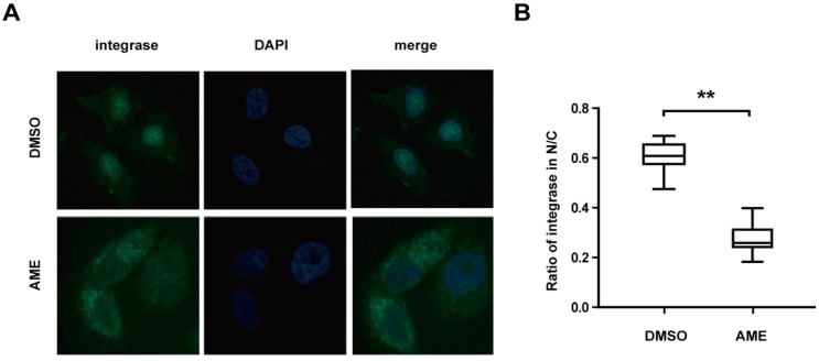
Figure 3.
AME blocks pre-integration complex (PIC) nuclear import. (A) HeLa cells were infected with VSV-G pseudotyped NL4-3Luc R-E- viruses in the presence of 60 µM AME or DMSO. Twelve h post-infection, cells were fixed and labelled with mouse anti-integrase (anti-IN) antibody followed by FITC-conjugated goat anti-mouse secondary antibody, and the nuclei were stained with 4′6′-diamidino-2-phenylindole (DAPI). Cells were analyzed by confocal microscopy (with a 60× objective lens); (B) Statistical analysis for the ratio of integrase in nuclei and cell cytosol. Ratios of fluorescence in nucleus and whole cells at 488 nm were measured from 25 DMSO-treated cells and 25 AME-treated cells randomly selected from three independent experiments. The fluorescence density at 488 nm was quantified by ImageJ and statistical analysis was performed in Graphpad Prism 5.0 software. The data represent the percentage of PIC in the nucleus. Error bars represent standard deviation (SD) of all cells in each group. ** p < 0.01 (determined by Student’s t test).

