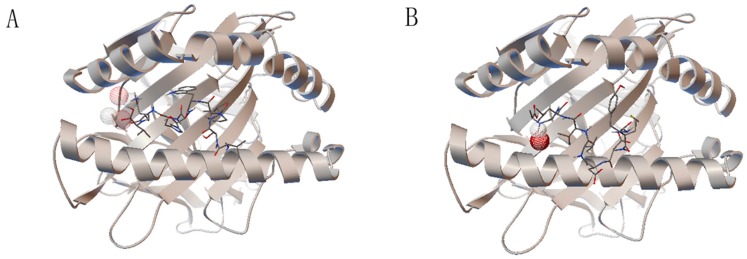
Figure 3.
The molecule docking result. The HLA-A0201 molecule is shown by secondary structure and the epitopes bind to the groove between the α1 and α2 domains of the HLA-A0201 molecule. (A) The epitope VSIPWTHKV has two hydrogen bonds, to lysine 8 and lysine 146 of the HLA-A0201 molecule; (B) epitope YMDDVVLGA has only one hydrogen bond, to alanine 9 of the HLA-A0201 molecule. The hydrogen bonds were shown by wireframe.

