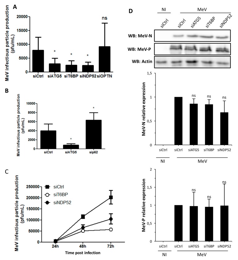
Figure 2.
Involvement of autophagy receptors in measles virus (MeV) replication. (A,B) HeLa cells were transfected with the indicated siRNAs for 48 h, then infected with MeV (multiplicity of infection (MOI) 0.1). 48 h post infection infectious virus particles were titrated by a plaque assay; (C) HeLa cells were transfected with the indicated siRNA for 48 h, then infected with MeV (MOI 1). One, two, or three days post infection, infectious virus particles were titrated by a plaque assay; (D) Cells were treated as in (A). Expression of measles virus N and P proteins was assessed by Western blotting. Representative results are shown and are accompanied by a graph representing the intensity of MeV-N and MeV-P expression over Actin normalized to Control condition.(A,B,D error bars and mean ± SD are from three independent experiments; C is one experiment representative of two independent ones carried out in duplicates). NI: non infected

