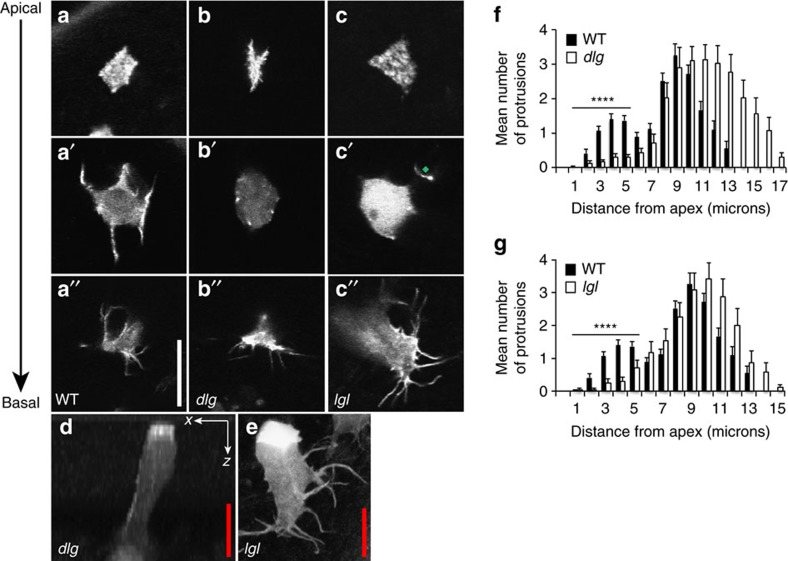
Figure 2. Loss of basolateral polarity proteins dlg or lgl leads to the specific loss of lateral protrusions.
(a–a″) Live imaging of a GFP:Moe-labelled wild-type epithelial cell showing apical (a) intermediate (a′) and basal (a″) confocal sections. (b,c) Apical, intermediate and basal confocal sections of GFP:Moe-labelled dlg (b–b″) and lgl (c–c″) mutant cells. Note the specific loss of protrusions in the intermediate sections. The green diamond highlights a protrusion that originates at a more basal plane. (d) An x–z confocal slice in a dlg mutant clone; (e) a z-projection of an lgl mutant cell, each again highlighting a lack of lateral protrusions. Note the abnormally thick and long basal protrusions in the lgl mutant cell (e). (f,g) Protrusion distributions for dlg (f) and lgl (g) mutant cells, highlighting a lack of intermediate level protrusions. Mutant cell distributions are represented by white bars, wild-type cell distributions by black bars. Error bars represent s.e.m. Wild-type n=50 cells from ten animals; dlg n=35 mutant cells from 19 animals; lgl n=40 mutant cells from 17 animals. Scale bars, 10 μm (white), 5 μm (red). Student's t-test was performed to determine statistical significance and P values are shown on graph. P>0.5 was considered not significant, *P<0.05, **P<0.01, ***P<0.001, ****P<0.0001.

