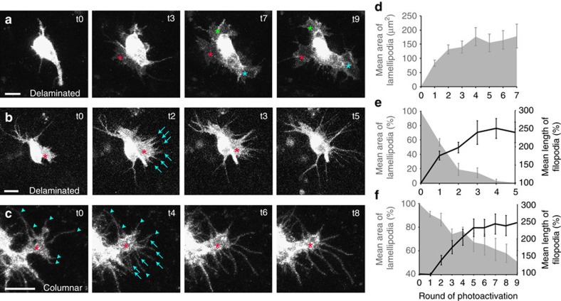
Figure 4. The level of Rac activity determines protrusion form.
(a–c) Live imaging of MARCM clones of epithelial cells overexpressing a constitutively active form of photoactivatable Rac, PA-RacQ61L (stills taken from Supplementary Movies S4–S6). (a) A delaminated epithelial cell rapidly develops three prominent lamellipodia upon photoactivation (asterisks). (b) A delaminated epithelial cell has a lamellipodium prior to photoactivation (asterisk). Upon photoactivation, the lamellipodium progressively retracts and new filopodia develop and grow (arrows). (c) A polarized epithelial cell within the epithelial sheet that possesses a basal lamellipodium (asterisk) as well as six pre-existing filopodial protrusions (arrowheads). Upon photoactivation, the lamellipodium slowly retracts and new filopodia develop and grow (arrows). (d–f) Quantification of mean lamellipodial area (grey area) and mean filopodial length (black line) over time, prior to photoactivation (t0) and after each consecutive round of photoactivation (t1–t9). (d) Delaminated cells with no lamellipodia prior to photoactivation (n=10 new lamellipodia following photoactivation). (e) Delaminated cells with prominent lamellipodia prior to photoactivation (n=8 lamellipodia, n=26 new filopodia following photoactivation). (f) Polarized epithelial cells within the epithelial sheet with prominent lamellipodia prior to photoactivation (n=12 lamellipodia, n=82 new filopodia following photoactivation). Error bars represent s.e.m. Scale bars, 10 μm.

