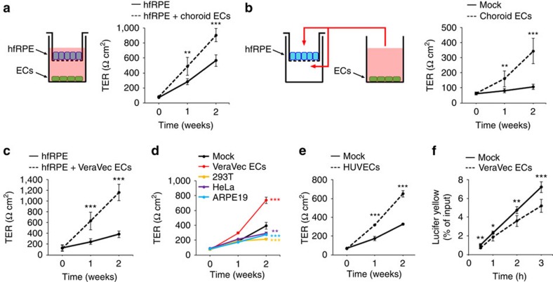
Figure 2. ECs increase TER and decrease paracellular permeability in hfRPE.
(a) Co-culture of hfRPE with choroid ECs as depicted promotes an increase in hfRPE TER (n=6, t-test). *hfRPE versus hfRPE+choroid ECs at each time point. (b) Conditioned media from choroid ECs promotes an increase in hfRPE TER (n=6, t-test). *mock versus choroid ECs at each time point. (c) Co-culture of hfRPE with VeraVec ECs as depicted in a promotes an increase in hfRPE TER (n=7, t-test). *hfRPE versus hfRPE+VeraVec ECs at each time point. (d) Conditioned media from VeraVec ECs, but not from 293T, HeLa or ARPE19 cells, promotes an increase in hfRPE TER (n=3, analysis of variance (ANOVA)). *mock versus VeraVec ECs, 293T, HeLa or ARPE19 at 2 weeks. (e) Conditioned media from naïve HUVECs promotes an increase in hfRPE TER (n=3, t-test). *mock versus HUVECs at each time point. (f) Incubation of hfRPE with conditioned media from VeraVec ECs for 2 weeks decreases Lucifer Yellow paracellular permeability across hfRPE (n=6, t-test). *mock versus VeraVec ECs at each time point. Data are presented as mean±s.d.

