Fig. 3.

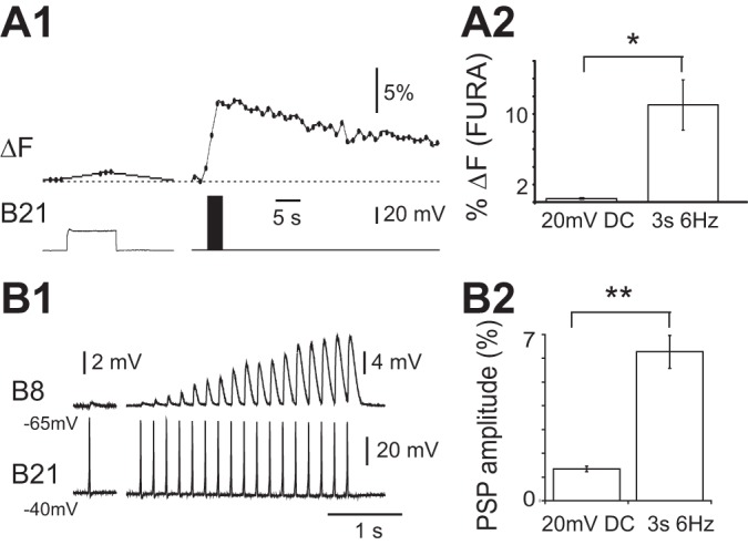
A1: spike-induced increases in calcium fluorescence in B21 vs. increases in calcium fluorescence (ΔF) induced by subthreshold depolarization. Ratiometric (fura) imaging was from the lateral process when B21 was maximally depolarized (i.e., depolarized by 20 mV; left) and when it was stimulated at 6 Hz for 3 s and minimally depolarized (i.e., depolarized by <10 mV; right). A2: group data for the experiment shown in A1. Note that spiking induced the larger signal (n = 5). B1: synaptic transmission between B21 and B8 under conditions where spike-induced increases in calcium fluorescence cannot summate (only one spike is triggered) vs. situation where B21 is repeatedly activated at a frequency where summation can occur. Left, B21 was centrally depolarized by 20 mV (i.e., was held at −40 mV) and a single spike induced. The PSP evoked in B8 was recorded with B8 at its normal resting potential (−65 mV). Right, repetitive stimulation of B21 at 6 Hz for 3 s with B21 and B8 at −40 and −65 mV, respectively. B2: group data for the experiment shown in B1. Note that with repetitive stimulation, B21-induced PSPs in B8 became progressively larger than the PSP potentiated by depolarization alone (n = 4). *P < 0.05; **P ≤ 0.01.
