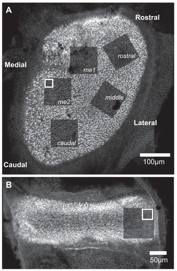
Fig. 2.

Topography-based confocal imaging of mouse vestibular epithelia. The locations of image stacks acquired from utricles and horizontal cristae are illustrated by the photobleached areas in specimens labeled with fluorophore-conjugated phalloidin. These appear as areas of diminished fluorescence after image stack acquisition. The white boxes represent the area of the 30-µm × 30-µm substack that was extracted from the larger image stack for the purpose of synapse counting. A: 5 regions of the mouse utricle were imaged for quantitative synapse analysis. These regions correspond to the utricular topography as follows: me1 and me2, medial extrastriola; caudal, caudal striola; middle, middle striola; and rostral, rostral striola. B: the planum of the horizontal crista (indicated by the absence of cruciate eminence featured in the superior and posterior cristae) was imaged for synapse quantification, illustrated by the postimaging photobleached area shown, and served as within-specimen control epithelium that is gravity insensitive.
