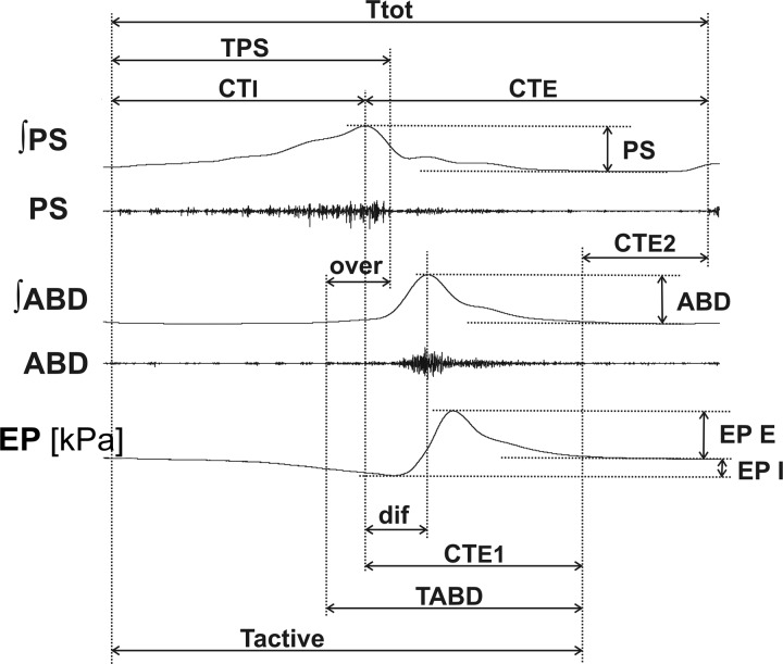
Fig. 2.
Illustration of cough measurements. ABD, abdominal muscle EMG; ʃABD, normalized moving average of the abdominal muscle EMG; CTE, duration of cough expiratory phase; CTE1, duration of active cough expiratory phase; CTE2, duration of quiescent period of cough expiration; CTI, duration of cough inspiratory phase; dif, time interval between peaks of parasternal and abdominal EMG activity; EP, esophageal pressure; EP E, EP I, expiratory and inspiratory esophageal pressure amplitudes, respectively; over, duration of parasternal and abdominal EMG coactivation; PS, parasternal muscle EMG; ʃPS, normalized moving average of parasternal muscle EMG; Tactive, duration of cough-related EMG activity; Ttot, total cough cycle duration; TABD, TPS, durations of abdominal and parasternal muscle discharge, respectively. Note that the decrementing part of the PS bursts (desI) corresponding to the interval TPS–CTI is not depicted.

