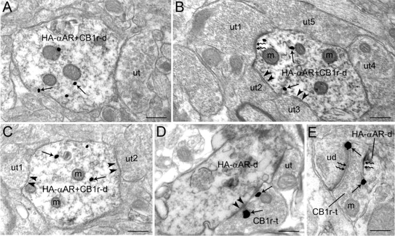
Figure 5.

Ultrastructural evidence for co-localization of HA-α-adrenoceptor and CB1 receptor in dendrites. A. An immunoperoxidase-labeled dendrite contains HA-α2A-adrenoceptor immunoreactivity as well as immunogold-silver labeling for CB1 receptor (HA-αAR+CB1r-d). This dendrite is directly contacted by an unlabeled terminal (ut). B. A dually labeled HA-αAR+CB1r dendrite receives an asymmetric type synapse (zigzag arrows) from an unlabeled terminal (ut1) and a symmetric type synapse (arrowheads) from ut2 and ut3. A fourth unlabeled terminal (ut4) directly contacts the HA-αAR+CB1r dendrite. C. A dually labeled HA-αAR+CB1r-d receives two symmetric type synapses (arrowheads) from ut1 and ut2. D. A CB1 receptor labeled axon terminal (CB1r-t) forms a symmetric type synapse (arrowheads) with HA-α-adrenoceptor labeled dendrite (HA-αAR-d). The same HA-αAR-d is contacted by an unlabeled terminal (ut). E. A CB1r-t forms an asymmetric type synapse (zigzag arrows) with an unlabeled dendrite (uD) and a HA-αAR-d. m: mitochondria; ut: unlabeled terminal. Scale bars, 0.5 μm.
